Click each image to open a higher resolution image. The materials included with the course include hundreds of pictures and numerous case studies just like these.
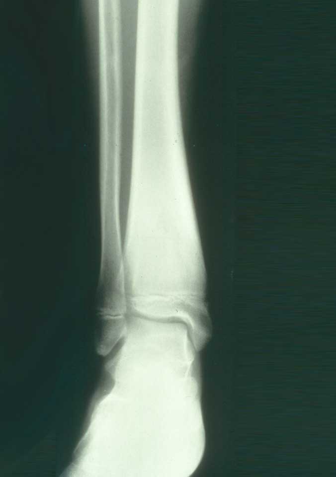
This 11 year old boy had a painful lesion of the distal right tibia. An excisional biopsy of the lesion was performed.
Where is the lesion?
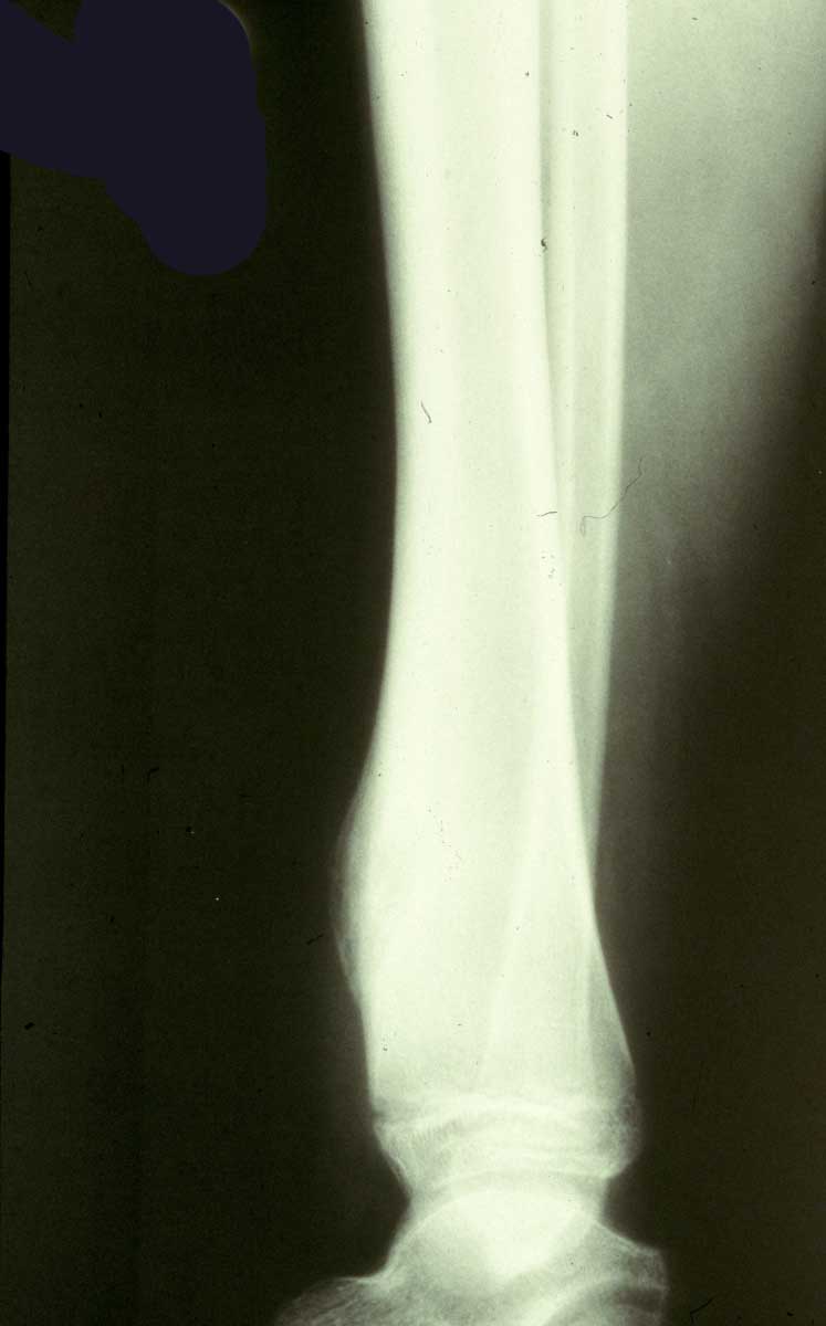
Describe the radiographic features of the lesion in this closer view.
Can you identify the cortex? What has happened to it? How did the changes occur? Do you see the lesion? What term do you apply to the central part of the lesion?
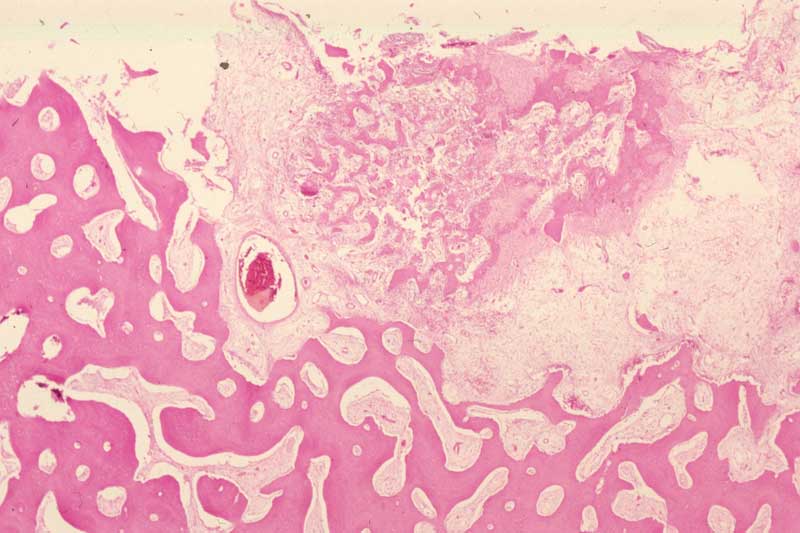
Describe the different histologic zones in this lesion.
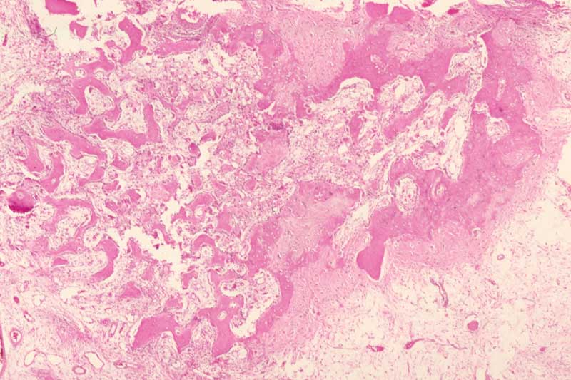
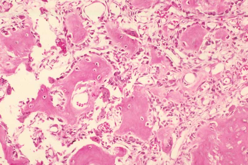
Identify the different cell types in this field. What type of bone is present? Describe the intertrabecular stroma; how is it different from normal bone marrow?
Name at least three lesions of bone that can have this histologic appearance. What causes the numerous osteoblasts to proliferate? (Note: there is a histologic clue in this field).