Click each image to open a higher resolution image. The materials included with the course include hundreds of pictures and numerous case studies just like these.
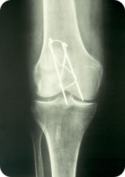
This 84 year old white female fractured her right patella and underwent an open reduction and internal fixation of the closed, displaced fracture. Abnormal synovial tissue was removed at surgery. Describe the radiographic findings.
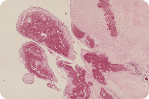
H&E with violet amorphous material. What type of tissue is involved by the deposits? Are the synovial lining cells normal?
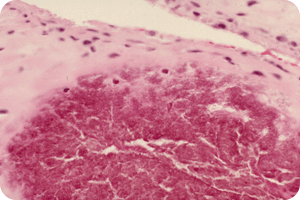
Close up view of the granular eosinophilic material. What is your differential diagnosis? What would you do to confirm your impression?
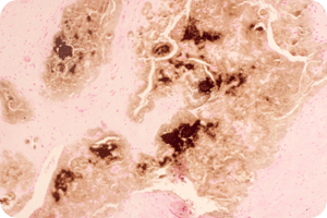
This is a Von Kossa stain for calcium (calcium stains dark brown to black). What does it show? Does this help you determine the final diagnosis?
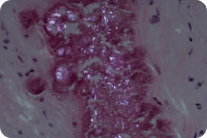
H&E section under polarized light. What are the small bright pink to white rectangular particles? What is your final diagnosis in this case?